Mammogram
A mammogram is a specialized medical imaging test that uses low-dose X-rays to take detailed pictures of the breast. It’s primarily used to screen for breast cancer and can often detect tumors or abnormalities that are too small to be felt during a physical exam.
There are two main types:
Screening mammogram: Done routinely in women without symptoms to check for early signs of breast cancer.
Diagnostic mammogram: Performed when there are symptoms (like a lump, pain, nipple discharge, or skin changes) or when something unusual is found on a screening mammogram.
During the test, the breast is gently compressed between two plates to spread out the tissue, which allows clearer images and reduces the amount of radiation needed.
It’s generally recommended that women begin regular screening mammograms around age 40–50, depending on personal risk factors and medical guidelines, and continue every 1–2 years.
Here’s what typically happens step by step during a mammogram so you know what to expect:
- Check-in and preparation
You’ll be asked to undress from the waist up and put on a gown that opens in the front.
You’ll need to remove jewelry or deodorant under your arms or on your breasts, since these can show up on the X-ray as white spots.
- Positioning
A technologist (usually female) positions you in front of the mammogram machine.
One breast at a time is placed on a flat plate.
- Compression
A second plate gently presses down on the breast.
This spreads out the tissue so the X-ray can capture a clearer image.
Compression only lasts a few seconds for each image.
It may feel uncomfortable or tight, but it should not be painful for most women.
- Imaging
Usually, two X-rays of each breast are taken:
Top-to-bottom view
Side view
Sometimes additional angles are taken if needed.
- Completion
The process usually takes about 20 minutes, though the actual compression only lasts a few seconds per picture.
Afterward, you can resume normal activities right away.
- Results
A radiologist reviews the images for signs of lumps, calcifications, or other changes.
Results are typically sent to you and your doctor within a few days.
Tips to make it easier:
Schedule your mammogram when your breasts are least tender (usually about a week after your period).
Wear a two-piece outfit so you only need to remove your top.
Let the technologist know if you have breast implants, are pregnant, or are breastfeeding.

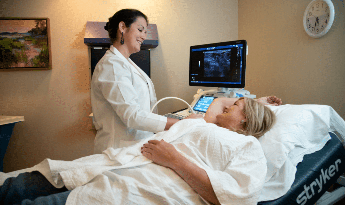
How we perform breast imaging
Preparation for ultrasound, stereotactic, or MRI guided breast biopsies: None needed for most patients. Because we use only local anesthesia, you can eat and drink as usual. Withholding blood thinners for a few days prior to a biopsy is suggested but not mandatory; check with your prescribing physician before stopping any medications.